Ear Reconstruction
Ear reconstruction is a two-stage procedure where the contours of the normal ear are shaped with costal cartilage and placed in a skin pocket to create the ear.
Model featured in photography
What can be achieved?
At Artiste Plastic Surgery Ear Reconstruction is performed for the following reasons:
- Microtia
- Craniofacial/hemifacial microsomia (CFM)
- Treacher Collins Syndrome (TCS)
- Goldenhar Syndrome
- Complex craniofacial conditions
- Other congenital anomalies
- After traumatic amputation
- After surgical excision
- After complications from Otoplasty surgery
Ear Reconstruction TECHNIQUES
Dr Jack Zoumaras uses the Francoise Firmin technique of ear reconstruction. This involves two stages. The first stage consists of harvesting rib cartilage and carving all the 3-D contours of the ear and the second stage involves creation of the post-auricular sulcus and elevation of the construct. Dr Jack Zoumaras spent 6 months in Paris with Francoise Firmin learning about ear reconstruction from the worlds best. In this time Dr Jack Zoumaras was trained in this technique and has already started a database of ear reconstructions performed in Sydney to date.
Microtia
Microtia is the congenital absence of the external ear/pinna. It affects 1 in 8000 live births and may be associated with other congenital syndromes such as CFM, TCS, and Goldenhar’s. It commonly affects the right side greater than the left and in some circumstances bilateral. It affects children of Asian descent slightly more than others. The cause of isolated Microtia is unknown and likely multi-factorial; there is a small genetic component with the risk of an affected parent transmitting to a child around 5%.
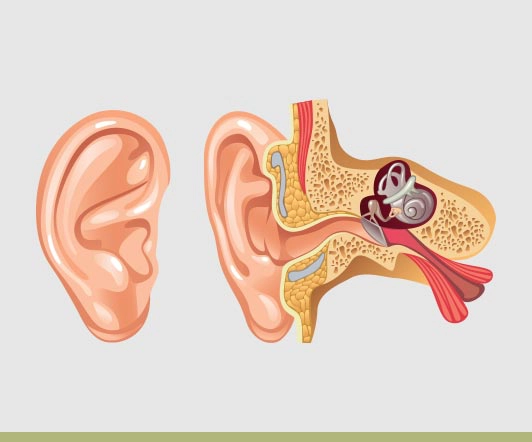
Depending upon the severity of the Microtia, the inner ear may or may not be developed. If not developed the child will not be able to hear from the ear. If the middle and inner ear is developed then conductive hearing through a bone anchored hearing device (BAHA) is possible. Patients with Microtia will be assessed before the age of 1 at a Microtia clinic with an audiologist, ENT and Plastic Surgeon and nurse co-ordinator.
The Francoise Firmin technique of ear reconstruction involves the formation of all contours of the ear including extra projection pieces during the first stage. As such more cartilage is required then other techniques and the surgery is performed from the age of 10 onwards. This produces superior aesthetic results. By the age of 9 it is possible to start considering reconstructing the external ear. At this age the ear is normally 80% adult size and the child’s rib cage has developed to the point that costal cartilage can be harvested for reconstruction. Most ear reconstructions are performed from the age of 12.
For more detailed information on Microtia management please see the following website microtia.com.au.
Other ear anomalies can be treated with similar techniques to Microtia reconstruction to form a normal looking ear.
Cryptotia:
Cryptotia is a congenital deformity where part of the ear is hidden within the scalp with no normal sulcus behind the ear.
Treatment consists of a series of flaps to create a sulcus and elevate the ear.
Constricted ears:
Constricted ears have all the structures of an ear but lack the size in both height and width. The constriction may be in the middle or upper part of the ear. After careful analysis of the normal ear or a parent’s ear in bilateral cases, a template is made which demonstrates the constriction of the ear and the loss in height and width. Treatment involves expanding the constricted ear to a normal size through use of cartilage grafts from the other ear or rib. The ear may also need to be set back.
Lop ear:
Is a constricted ear where the vertical height and width is reduced in size and results in folding of the scapha.
Cup Ear:
Is a lop ear that is also prominent.
Treatment consists off expanding the height/width of the ear through use of cartilage grafts from the rib or contralateral normal ear.
Prominent ears:
Discussed under Otoplasty
FAQs
The second stage is after 6 months to allow the new ear to heal into the pocket and be stable.
The second stage is overnight surgery and pain is minimal compared to the first stage. Again a head bandage is worn for a few days.
The second stage involves elevating the flat 3D construct of the ear from the ear pocket to create elevation and a post-auricular sulcus. A skin graft and/or temporoparietal fascia flap is sometimes needed to project the ear. This stage can be combined with surgery on the other ear to create symmetry.
You will remain in hospital for 4 days with a head bandage and suction drains that will be changed regularly. After discharge the drains will be removed and a lighter dressing applied. Most of the pain initially comes from the chest wall that is managed with intravenous and oral medications.
Rib cartilage is harvested from the same side as the ear to be reconstructed. After preparing the ear pocket the rib cartilage is sculpted to form a new ear. This sculpting is modelled after a template of the normal ear. The entire 3-D contour of the ear is reconstructed during this stage. The new ear is placed into the pocket with suction tubing. Due to a lack of skin the new ear is flat against the head.
As Seen On